28++ Facs buffer flow cytometry for Android Phone
Home » Wallpaper » 28++ Facs buffer flow cytometry for Android PhoneNews Facs buffer flow cytometry post are available in this site. Facs buffer flow cytometry are a topic that is being searched for and liked by netizens now. You can Get the Facs buffer flow cytometry files here. Download all free photos.
If you’re searching for facs buffer flow cytometry images information linked to the facs buffer flow cytometry interest, you have come to the ideal site. Our website always provides you with hints for viewing the highest quality video and image content, please kindly search and find more enlightening video content and images that match your interests.
Facs Buffer Flow Cytometry, Pharmingenstain buffer (bsa) is useful for the dilution and application of fluorescent reagents as well as for the suspension, washing, and storage of cells destined for flow cytometric analysis (or fluorescence microscopy). Perform fluorescence activated cell sorting (facs), or flow cytometric analysis. It is a useful scientific instrument, as it provides. Flow cytometry (direct immunofluorescence staining):
 Flow Cytometry Flow cytometry, Medical technology, Tech From pinterest.com
Bill eades and the university of rochester medical center flow cytometry shared resource share resource direct dr. The buffer can be simplified to hbss with 1% fbs. Flow cytometry permits the detection of transcription factors within discrete immune cell subsets among a heterogeneous population and provides a sensitive approach to analyzing an immune response. Perform fluorescence activated cell sorting (facs), or flow cytometric analysis. Ensure that you ph your buffer properly.
Bring a few mls of additional sorting buffer in case we need to dilute the sample.
Alternatively, samples can be fixed with 2% paraformaldehyde fixation buffer and stored at 4°c in the dark for up to one week before flow cytometry analysis. Large differences in mean channel values for fsc and ssc between major leukocyte populations 3. Timothy bushnell, ph d for their review and input on this technical document. Use the buffer that is best for your cells: Store at 4°c in darkness and acquire preferably within 24 hours. Low cvs on fsc and ssc 2. Due to presence of bsa or other proteins of fbs cells get stable and less sensitive to damage.
 Source: pinterest.com
Flow Cytometry Flow cytometry, Medical technology, Tech Our flow cytometry staining buffer is designed for use in immunofluorescent staining protocols of cells in suspension. Bill eades and the university of rochester medical center flow cytometry shared resource share resource direct dr. *do not add sodium azide to buffers if you are concerned with recovering cell function e.g. The buffer can be simplified to hbss with 1% fbs. Ion chelating drugs, azt, etc.) are known to interfere with this lysis procedure. Bring a few mls of additional sorting buffer in case we need to dilute the sample.
 Source: pinterest.com
MACS Flow Cytometry offers customized highthroughput The samples should be resuspended in cell staining buffer. Large differences in mean channel values for fsc and ssc between major leukocyte populations 3. It is a useful scientific instrument, as it provides. Ensure that you ph your buffer properly. Beyond antibody reagents, flow cytometry requires the right types of buffers for optimal staining. People use �protein containing buffers� for flow cytometry is to prevent cells from sticking to the side of plastic tubes (or other culture labware) as well as preventing cell clumping.
 Source: pinterest.com
Global Flow Cytometry Market Industry Trends and Flow cytometry (direct immunofluorescence staining): Neonatal rbcs are extremely resistant to osmotic stress/lysis and some drugs (i.e. Beyond antibody reagents, flow cytometry requires the right types of buffers for optimal staining. The samples should be resuspended in cell staining buffer. Flow cytometer and its tubing from clogging up. Flow cytometry permits the detection of transcription factors within discrete immune cell subsets among a heterogeneous population and provides a sensitive approach to analyzing an immune response.
 Source: pinterest.com
UCFlow Flow Cytometry news, reviews, and tips. A First Flow cytometer and its tubing from clogging up. Effect of various lysing methods and reagents on flow cytometry immunophenotyping 2 1. *do not add sodium azide to buffers if you are concerned with recovering cell function e.g. Phosphate buffered saline (pbs) is a common suspension buffer. Our flow cytometry staining buffer is designed for use in immunofluorescent staining protocols of cells in suspension. Timothy bushnell, ph d for their review and input on this technical document.
 Source: pinterest.com
BD FACS Analyzer (With images) Flow cytometry Impact on the stability of tandem fluorochromes 6. Preservation of fluorochrome brightness 5. It provides a method for sorting a heterogeneous mixture of biological cells into two or more containers, one cell at a time, based upon the specific light scattering and fluorescent characteristics of each cell. Flow cytometry (direct immunofluorescence staining): Use the buffer that is best for your cells: This convenient list separates out flow cytometry applications by their intended target.
 Source: pinterest.com
Using centrifugal elutriation and flow cytometry to answer Impact on the stability of tandem fluorochromes 6. This flow cytometry staining buffer is a buffered saline solution containing fetal bovine serum and sodium azide (0.09%) as a preservative. Phosphate buffered saline (pbs) is a common suspension buffer. Impact on the stability of tandem fluorochromes 6. It provides a method for sorting a heterogeneous mixture of biological cells into two or more containers, one cell at a time, based upon the specific light scattering and fluorescent characteristics of each cell. Another reason that people use �protein containing buffers� for flow cytometry is to prevent cells from sticking to the side of plastic tubes (or other culture labware) as well as preventing cell clumping.
 Source: pinterest.com
Figure 2 Flow cytometric analysis of the cellular uptake Flow cytometry permits the detection of transcription factors within discrete immune cell subsets among a heterogeneous population and provides a sensitive approach to analyzing an immune response. Large differences in mean channel values for fsc and ssc between major leukocyte populations 3. *do not add sodium azide to buffers if you are concerned with recovering cell function e.g. Run samples immediately on the flow cytometer (take them down to cytometry). The buffer can be simplified to hbss with 1% fbs. Refer to the following section on intracellular staining buffers prior to any transcription factor staining analysis.
 Source: pinterest.com
Our Synaptic Marker being used to stain tissue from space Pharmingenstain buffer (bsa) is useful for the dilution and application of fluorescent reagents as well as for the suspension, washing, and storage of cells destined for flow cytometric analysis (or fluorescence microscopy). Perform fluorescence activated cell sorting (facs), or flow cytometric analysis. This flow cytometry staining buffer is a buffered saline solution containing fetal bovine serum and sodium azide (0.09%) as a preservative. Refer to the following section on intracellular staining buffers prior to any transcription factor staining analysis. Flow cytometry (direct immunofluorescence staining): Special acknowledgment and thanks to the siteman cancer flow cytometry core facility core director mr.
 Source: pinterest.com
Flow cytometry data can be visualized in a dot plot, where Refer to the following section on intracellular staining buffers prior to any transcription factor staining analysis. Add the optimized dilution of antibodies to the respective tubes and incubate at 4°c (on ice) for 30 minutes in the dark. People use �protein containing buffers� for flow cytometry is to prevent cells from sticking to the side of plastic tubes (or other culture labware) as well as preventing cell clumping. Preservation of fluorochrome brightness 5. Wash the cells twice in cold stain buffer (fbs) and pellet the cells by centrifugation (e.g., 300 x g at 4°c). Neonatal rbcs are extremely resistant to osmotic stress/lysis and some drugs (i.e.
 Source: pinterest.com
Pin by CogniBrain on Study in 2020 Study, Flow cytometry Use the buffer that is best for your cells: Bill eades and the university of rochester medical center flow cytometry shared resource share resource direct dr. Timothy bushnell, ph d for their review and input on this technical document. Impact on the stability of tandem fluorochromes 6. It is a useful scientific instrument, as it provides. Our flow cytometry staining buffer is designed for use in immunofluorescent staining protocols of cells in suspension.
 Source: pinterest.com
Preclinical & Clinical Studies Using Flow Cytometry Use the buffer that is best for your cells: Bill eades and the university of rochester medical center flow cytometry shared resource share resource direct dr. Our flow cytometry staining buffer is designed for use in immunofluorescent staining protocols of cells in suspension. The samples should be resuspended in cell staining buffer. Run samples immediately on the flow cytometer (take them down to cytometry). Special acknowledgment and thanks to the siteman cancer flow cytometry core facility core director mr.
 Source: pinterest.com
Running a Basic 2 color Flow Cytometry Experiment in BD Basic flow cytometry staining protocol written by: This buffer can be used for antibody and Use the buffer that is best for your cells: Ion chelating drugs, azt, etc.) are known to interfere with this lysis procedure. The buffer can be simplified to hbss with 1% fbs. Pharmingenstain buffer (bsa) is useful for the dilution and application of fluorescent reagents as well as for the suspension, washing, and storage of cells destined for flow cytometric analysis (or fluorescence microscopy).
 Source: pinterest.com
Image result for flow cytometry Imunologia Run samples immediately on the flow cytometer (take them down to cytometry). Another reason that people use �protein containing buffers� for flow cytometry is to prevent cells from sticking to the side of plastic tubes (or other culture labware) as well as preventing cell clumping. Ensure that you ph your buffer properly. The purpose of the azide in these buffers is to prevent microbial growth, but these buffers are used so quickly (and are extremely cheap to make) that you shouldn�t run into any problems. Place samples in 12 x 75 mm falcon® tubes and analyze by flow cytometry as soon as possible (within 1 hour). Store at 4°c in darkness and acquire preferably within 24 hours.
 Source: pinterest.com
Figure 9 Confocal fluorescence microscopy images and flow Flow cytometry permits the detection of transcription factors within discrete immune cell subsets among a heterogeneous population and provides a sensitive approach to analyzing an immune response. This buffer can be used for antibody and Special acknowledgment and thanks to the siteman cancer flow cytometry core facility core director mr. Beyond antibody reagents, flow cytometry requires the right types of buffers for optimal staining. Perform fluorescence activated cell sorting (facs), or flow cytometric analysis. Neonatal rbcs are extremely resistant to osmotic stress/lysis and some drugs (i.e.
 Source: pinterest.com
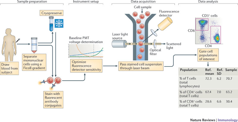
This shows the general scheme of flow cytometry Flow This buffer can be used for antibody and Basic flow cytometry staining protocol written by: Wash the cells twice in cold stain buffer (fbs) and pellet the cells by centrifugation (e.g., 300 x g at 4°c). Add the optimized dilution of antibodies to the respective tubes and incubate at 4°c (on ice) for 30 minutes in the dark. This flow cytometry staining buffer is a buffered saline solution containing fetal bovine serum and sodium azide (0.09%) as a preservative. Neonatal rbcs are extremely resistant to osmotic stress/lysis and some drugs (i.e.
 Source: in.pinterest.com
BioLegend offers an extensive selection of antibody Preservation of fluorochrome brightness 5. The buffer can be simplified to hbss with 1% fbs. Run samples immediately on the flow cytometer (take them down to cytometry). Preservation of fluorochrome brightness 5. Wash the cells twice in cold stain buffer (fbs) and pellet the cells by centrifugation (e.g., 300 x g at 4°c). Place samples in 12 x 75 mm falcon® tubes and analyze by flow cytometry as soon as possible (within 1 hour).
 Source: pinterest.com
Hematology and FlowCytometry Analyzers and Reagents Notes dnase i requires a concentration of at least 1 mm magnesium to work effectively, although 5 mm is optimal. The additional cations in the recipe promote better viability. Special acknowledgment and thanks to the siteman cancer flow cytometry core facility core director mr. Flow cytometer and its tubing from clogging up. This convenient list separates out flow cytometry applications by their intended target. Beyond antibody reagents, flow cytometry requires the right types of buffers for optimal staining.
 Source: pinterest.com
All Cartoons Cell, Science humor Timothy bushnell, ph d for their review and input on this technical document. Perform fluorescence activated cell sorting (facs), or flow cytometric analysis. This flow cytometry staining buffer is a buffered saline solution containing fetal bovine serum and sodium azide (0.09%) as a preservative. Effect of various lysing methods and reagents on flow cytometry immunophenotyping 2 1. It is a useful scientific instrument, as it provides. Preservation of fluorochrome brightness 5.
If you find this site beneficial, please support us by sharing this posts to your preference social media accounts like Facebook, Instagram and so on or you can also bookmark this blog page with the title facs buffer flow cytometry by using Ctrl + D for devices a laptop with a Windows operating system or Command + D for laptops with an Apple operating system. If you use a smartphone, you can also use the drawer menu of the browser you are using. Whether it’s a Windows, Mac, iOS or Android operating system, you will still be able to bookmark this website.
This site is an open community for users to submit their favorite wallpapers on the internet, all images or pictures in this website are for personal wallpaper use only, it is stricly prohibited to use this wallpaper for commercial purposes, if you are the author and find this image is shared without your permission, please kindly raise a DMCA report to Us.
Category
Related By Category
- 26+ Blue hibiscus flower tattoo meaning for Desktop Background
- 24++ Flower hair band images for Homescreen
- 37+ Data flow diagram examples in software engineering for Desktop Background
- 17++ Furnace air flow switch for Desktop Background
- 45++ Flow past tense meaning for Windows Mobile
- 44+ Anemone flower meaning betrayal for Android Phone
- 25+ Flower mandala coloring pages for adults for Homescreen
- 17++ Artificial flower pot price for Homescreen
- 22+ Black rose flower price for Desktop Background
- 33+ Cute flower canvas paintings for Desktop Background
